Above:
Building upon the success of NASA’s Mars Science Laboratory mission involving the Curiosity rover, the Mars 2020 rover mission will look carefully at Martian rocks to address additional high-priority science goals for Mars exploration, including key questions about the potential for life on the Red Planet and select samples for later return to Earth.
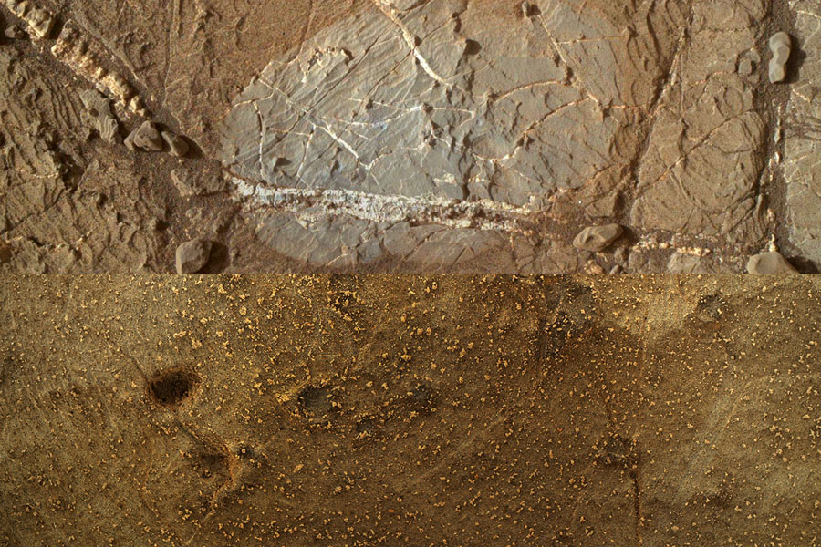
PIXL
X-Ray maps of Martian Rock Chemistry
Daniel Wilson
Led by JPL, Mars 2020 takes the next step in NASA’s Mars Exploration Program, probing the Martian rocks for evidence of past life. The four goals of Mars 2020 are to identify past environments capable of supporting microbial life, seek signs of biosignatures for past microbial life in those habitable environments, collect core rock and “soil” samples and store them on the Martian surface, and test oxygen production from the Martian atmosphere.
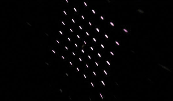
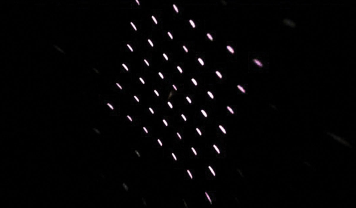
The Planetary Instrument for X-ray Lithochemistry (PIXL) for the Mars-2020 rover is an X-ray fluorescence instrument that rapidly measures elemental chemistry at sub-millimeter scales by focusing an X-ray beam to a tiny spot on the target rock or soil and analyzing the induced X-ray fluorescence. The PIXL Optical Fiducial Subsystem (OFS) uses two structured-light illuminators with a camera system to localize the instrument relative to the sample, and to map the topography of the surrounding area.
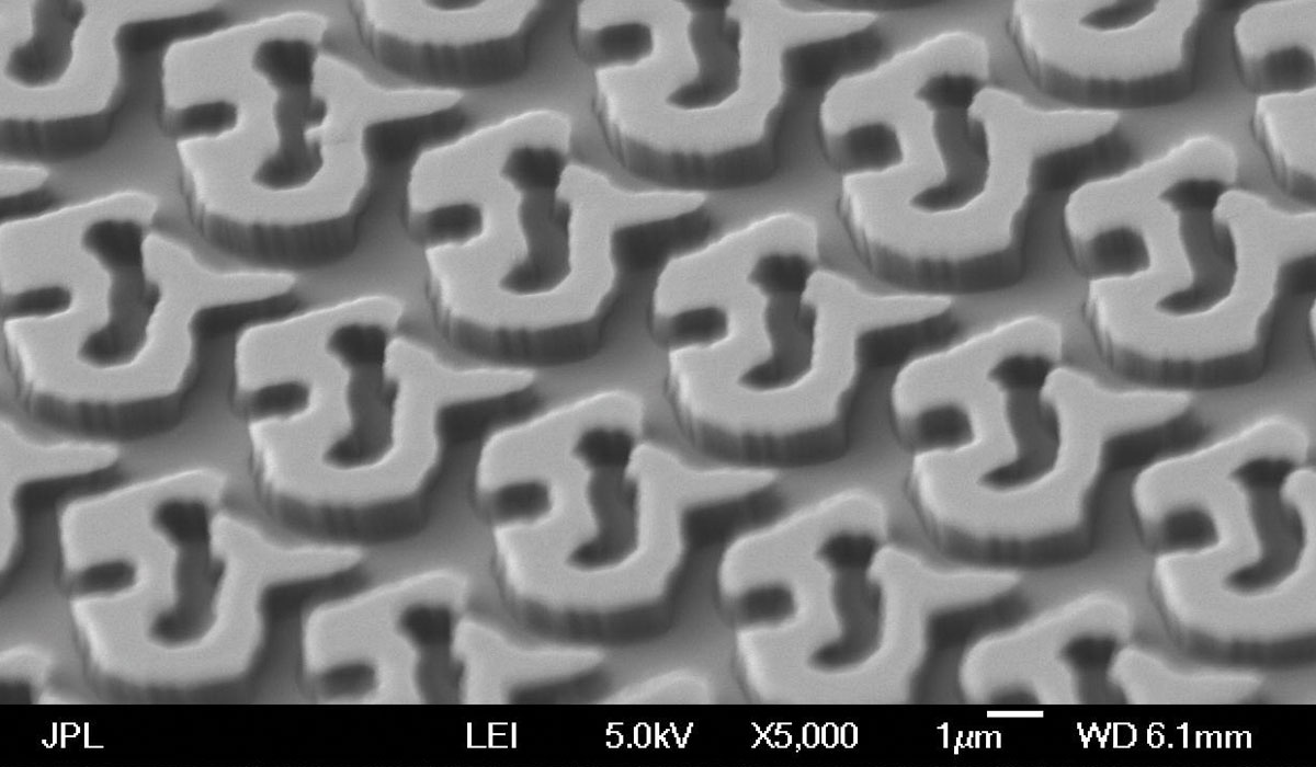
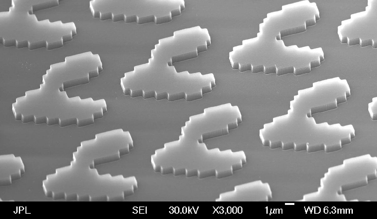
Critical to the OFS operation are diffractive spot array generators that produce 3x5 and 7x7 patterns of laser spots. The spot array generators are transmissive two-dimensional computer-generated hologram (CGH) gratings that are designed, electron-beam patterned, and plasma etched into fused silica substrates in the MDL. Figure 1 shows the design and a microscope image of the fabricated 7x7 CGH.
Each cell of the CGH grating is made up of 160 nm square pixels and when etched to the proper depth in fused silica, the surface relief imparts a phase pattern on the laser beam (830 nm wavelength), causing it to diffract into 7x7 spots with near equal intensity.
Gratings for three-by-five and a seven-by-seven spot arrays have been fabricated and two of each type were delivered to the project. They have been mounted in the OFS and successfully tested.
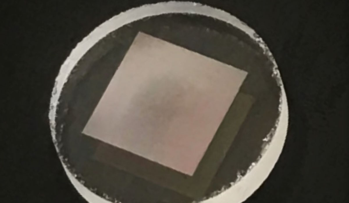
Seven-by-seven spot array seen through the above grating.
+ Larger image
Scanning electron microscope image of the seven-by-seven spot array grating.
+ Larger image
Scanning electron microscope image of the three-by-three spot array grating.
+ Larger image


